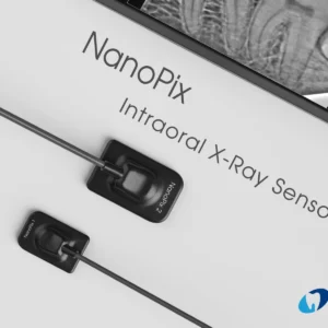
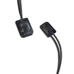
UNICORN - Xpect Vision XVD2530 RVG
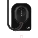
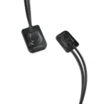
Xpect Vision XVD2530 RVG, a groundbreaking advancement in digital radiography that redefines the boundaries of dental imaging.
₹51,785.00
Description
Xpect Vision XVD2530 RVG, a groundbreaking advancement in digital radiography that redefines the boundaries of dental imaging. Utilizing Photon Counting Detection (PCD) technology, this intraoral sensor captures immaculate digital images with unparalleled clarity and efficiency. In just three seconds, dentists can obtain high-quality diagnostic images boasting exceptional dynamic range and contrast resolution, facilitating precise treatment planning and monitoring. Moreover, its durable casing withstands shocks, bites, and drops, while its waterproof design ensures optimal hygiene and safety. Compatible with existing imaging technologies and supporting lightning-fast data transmission via USB connection, the Xpect Vision RVG seamlessly integrates into any dental practice, enhancing workflow efficiency and patient care.
FEATURES:-
Xpect Vision XVD2530 RVG
Higher sensitivity intraoral sensor helps in easy capturing of images.
APS CMOS technology-based intraoral sensor ensures high-quality image.
Ergonomically designed with rounded edges helps in easy positioning in patients.
Highly durable & waterproof sensor with strong tear-resistant.
Convenient operation with the new Runyes software.
Various image processing tools provide a diagnosable image for treatment planning.
Multi-user solution, easy connection to PACs system under DICOM 3.0 protocol
KEY SPECIFICATION:-
Technology: PCD
Gray Scale: 16 Bits, Ultra High Scale (Highest in the industry)
Image Resolution: 14LP/MM
Protection : IP68
Size: 1.5
Connection: USB connection/ Wireless
Connection Cable: 3m (Extendable)
Support System: Windows 7, 8, and 10
Dimension of active area: 25 X 30 Mm
Sensor plate thickness: 5mm
DIRECTION OF USE:-
Prepare the Patient: Ensure the patient is positioned comfortably in the dental chair. Place a protective cover on the sensor for infection control.
Connect the Sensor: Connect the Runyes Intraoral Sensor to the USB 2.0 high-speed port on your computer.
Position the Sensor: Use the unique size of 1.3 to easily position the sensor inside the patient’s mouth. Follow proper placement guidelines for optimal imaging.
Capture Images: Open the imaging software on your computer. Select the patient and imaging settings. Instruct the patient to stay still and capture images using the sensor.
Review and Save Images: Review the captured images for quality and diagnostic accuracy. Save the images to the patient’s digital record.
Related products
-
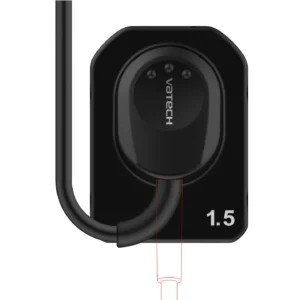
Vatech RVG EZ Sensor Classic Size 1.5, 5 year warranty
₹102,678.00Add to WishlistAdd to Wishlist -
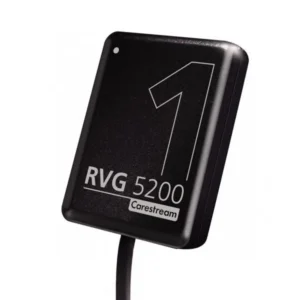
Carestream Kodak Rvg
₹99,553.00Add to WishlistAdd to Wishlist -
Eighteeth Nanopix Dental RVG
₹66,071.00 – ₹76,071.00Add to WishlistAdd to Wishlist -
Woodpecker RVG i Sensor Size Xpect Vision XVD2530 RVG
₹55,357.00 – ₹62,500.00Add to WishlistAdd to Wishlist

